Anatomy of the Nose: A Surgeon's Guide
The parts of the nose explained: bones, cartilages, septum, turbinates and valves, and what each one means for how your nose looks and breathes
I operate on the structures on this page every week. This is the anatomy of the nose as I explain it to my own patients: clear enough to follow without a medical degree, accurate enough to be worth your time, and connected at every step to the clinical questions that actually matter, from blocked breathing to what rhinoplasty can and cannot change.
Written and reviewed by Mr David Whitehead, Consultant ENT & Facial Plastic Surgeon, GMC 4372358. Last updated .
The Short Version
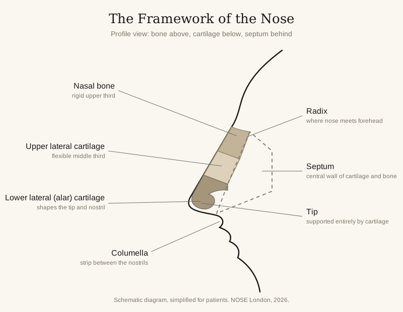
The external nose is a framework of cartilage and bone under the skin: two nasal bones form the rigid upper third, and the upper and lower lateral cartilages form the flexible lower two thirds, including the tip. Inside, the nasal cavity is divided in two by the septum; three pairs of turbinates on each side wall condition the air you breathe; the nasal valves are the narrow segments that limit airflow; and the paranasal sinuses drain into the whole system. The main functions of the nose include breathing, filtering and humidifying air, smell, and shaping the voice. Nearly every common nose problem, and nearly everything rhinoplasty does, comes down to one of these few structures.
I am Mr David Whitehead, a consultant ENT and facial plastic surgeon at NOSE London. Anatomy is not an academic detour on this website: it is the working knowledge behind every septoplasty and rhinoplasty I perform, and it matters for an important, practical reason: patients who understand their own anatomy make better decisions. The sections below take the nose from the outside in.
The External Nose: What You Can See and Feel
Surgeons think of the external nose in thirds, and once you know the rule you can feel it on your own face. The upper third is bone. The lower two thirds are cartilage. That single fact explains most of what goes wrong with noses, and most of what rhinoplasty does.
- The nasal bones: two small, paired bones forming the rigid bridge. They start at the radix, the dip where the nose meets the forehead, and they are what breaks in a nasal fracture.
- The upper lateral cartilages: the flexible middle third, fused to the underside of the nasal bones and to the septum in the midline. Press halfway down your nose and feel the give.
- The lower lateral cartilages (the alar cartilages): two curved springs that form the nasal tip and the rim of each nostril. Their size, strength and angle largely decide how the tip looks: boxy, bulbous, drooping or refined.
- The columella: the strip of skin and cartilage between the nostrils, running from the tip to the upper lip.
- The skin envelope: the soft tissue draped over the whole framework. Skin thickness varies enormously between people and it matters: thick skin hides fine cartilage work, thin skin reveals every contour beneath it.
The shape you see in the mirror is simply this framework of cartilage and bone, wrapped in your particular skin. Every nose shape I describe on my nose shapes pages traces back to some combination of these few structures.
Inside the Nose: The Nasal Cavity
Behind the nostrils, the nasal cavity runs surprisingly deep: about seven centimetres straight back to the top of the throat, divided down the middle into two airways by the septum.
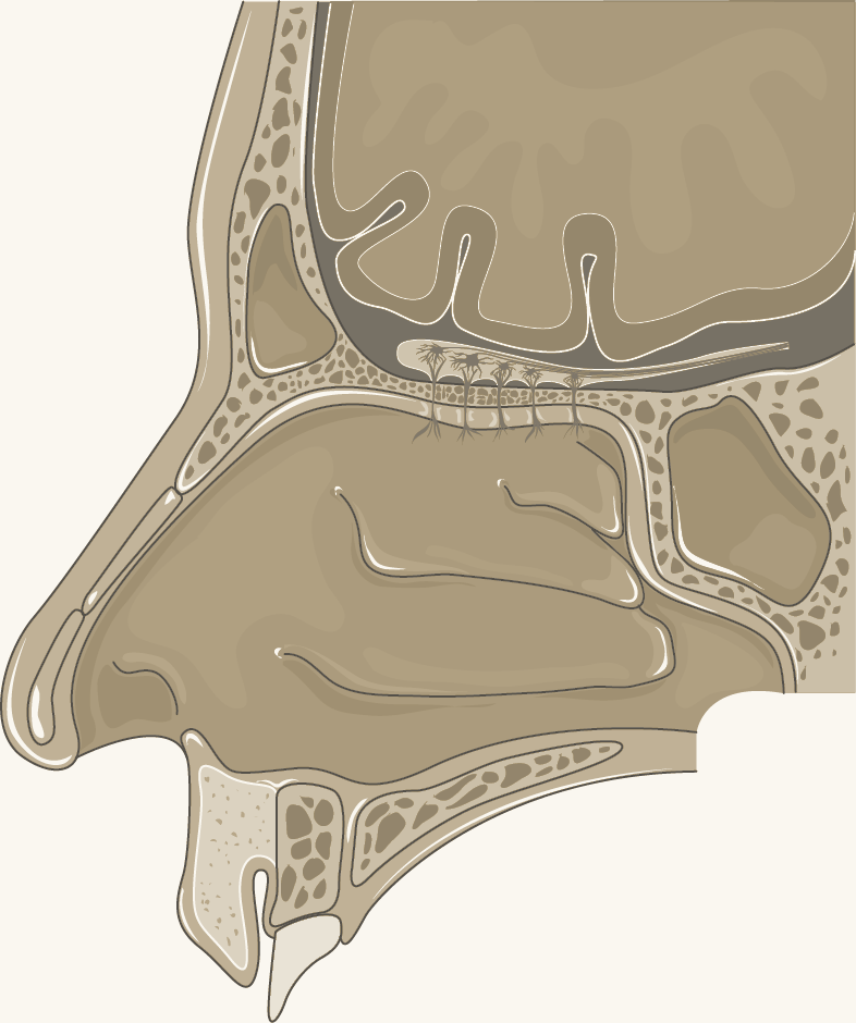
The nasal cavity in cross-section: the scroll-shaped turbinates on the side wall, the palate below, the skull base and brain above. Adapted (recoloured) from Servier Medical Art, CC BY 4.0.
The nasal septum is the central wall. The quadrangular cartilage forms the front, and two bones, the perpendicular plate of the ethmoid bone and the vomer, form the back. A perfectly straight septum is rare; a septum deviated enough to block breathing is one of the most common problems I treat, and correcting it is called septoplasty.
The turbinates are the structures patients have usually never heard of until one enlarges. They are three pairs of scroll-shaped shelves on the lateral nasal wall (the side wall of each airway): the inferior, middle and superior turbinates. Covered in a thick blanket of warm, wet lining, their role is to condition air on its way to the lungs: warming it, humidifying it and filtering it. The channels beneath them, each called a meatus, are where the sinuses and the tear duct drain. When the lining swells, with a cold, with allergy, or as part of the normal nasal cycle, the airway narrows and the nose feels blocked.
The nasal valves are the pinch points. The internal nasal valve, the narrow angle between the septum and the upper lateral cartilage, is the tightest part of the entire airway; the external nasal valve is the nostril opening itself. Because they are the narrowest segments, small changes here have a large effect on airflow, which is why valve collapse makes breathing so difficult and why I assess both valves in every patient.
High in the roof of the cavity, just beneath a thin sheet of bone called the cribriform plate, sits the olfactory nerve: the nerve of the sense of smell. Air swirling up to this area is what lets you smell, which is why a badly blocked nose blunts both smell and, because flavour is mostly smell, taste.
The Paranasal Sinuses
The sinuses are air-filled chambers in the bones of the face and skull, connected to the nasal cavity by narrow drainage channels. There are four pairs: the maxillary sinus in each cheek (the largest), the frontal sinuses in the forehead, the ethmoid sinuses between the eyes, and the sphenoid sinuses deep behind them.
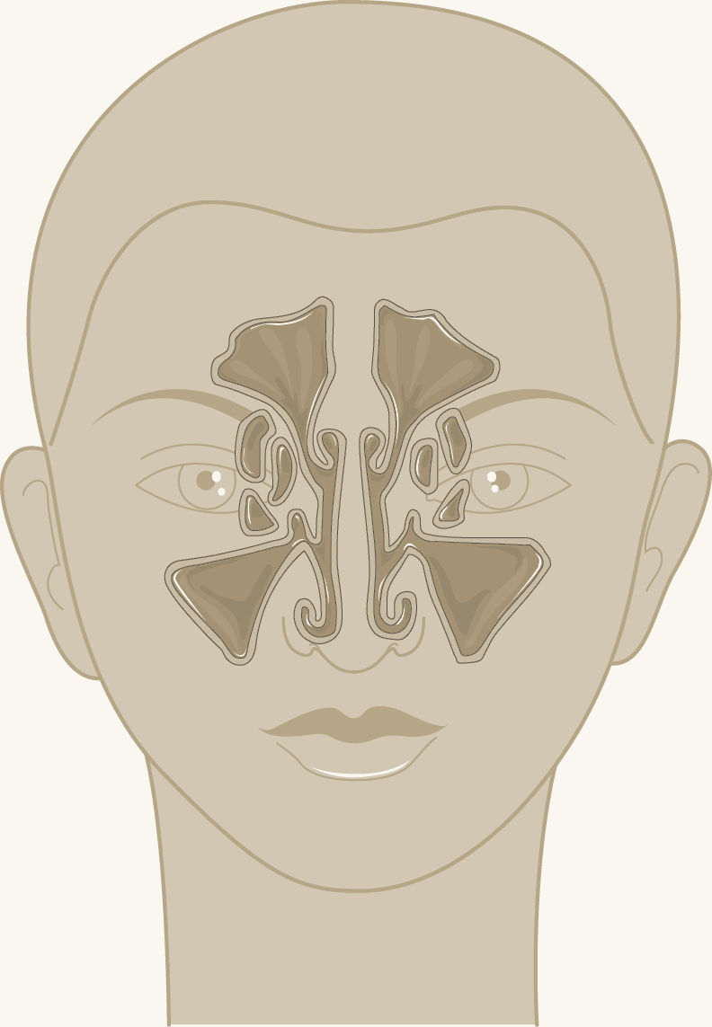
Where the sinuses sit: frontal in the forehead, ethmoid between the eyes, maxillary in the cheeks. Adapted (recoloured) from Servier Medical Art, CC BY 4.0.
Most drain into the middle meatus, under the middle turbinate, which makes that small area the busiest junction in the nose. Anything that obstructs it, swelling, a deviated septum, or nasal polyps, can trap mucus in the sinuses and set up the pressure, congestion and infection cycle patients know as sinusitis.
The sinuses also lighten the skull and add resonance to the voice, which is why you sound different when congested. As an ENT surgeon I treat the nose and sinuses as one connected system, because anatomically that is exactly what they are: the nose is the front door of the whole upper respiratory tract.
Blood Supply and Nerves
The nose has one of the richest blood supplies of any structure in the head and neck, fed from several arteries at once: the anterior ethmoidal artery from above (a branch of the ophthalmic artery), the sphenopalatine and greater palatine arteries from behind, and the superior labial artery from the lip below.
These vessels meet in an area on the front of the septum called Little's area, and that anatomical detail has a practical consequence every parent knows: it is where the great majority of nosebleeds start. Clinicians divide nosebleeds into anterior and posterior bleeds: anterior bleeds from Little's area are the common, manageable kind, while the rarer posterior bleed, from deeper vessels in older patients, is the kind that needs hospital care.
The generous blood supply is also good news for surgery: it is one reason the nose heals so reliably after rhinoplasty, and it is what allows surgeons to lift the skin safely from the framework during an operation. Sensation comes from branches of the trigeminal nerve, which is why a fresh injury to the nose brings tears to your eyes, and why the tip can feel temporarily numb after surgery while those fine nerve branches recover.
Bone or Cartilage? What the Nose Is Made Of
Patients ask this constantly, and the answer is: both, in a precise arrangement. The upper third of the nose is bone. The lower two thirds, everything you can wiggle, is cartilage. The septum inside follows the same pattern: cartilage at the front, bone at the back.
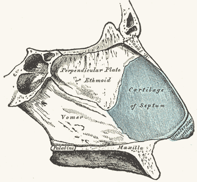
The septum's construction, from Gray's Anatomy (1918, public domain): the cartilage of the septum in front, the perpendicular plate of the ethmoid and the vomer behind. The century-old labels match the terms surgeons still use today.
The distinction matters clinically. Bone breaks: that is the fracture you get from a football or a fall, and it shows the classic bruising and the bent bridge. Cartilage bends, buckles and, over a lifetime, weakens: the tip of the nose drops slightly with age not because the nose keeps growing, but because the cartilages soften and the skin thickens and stretches. Cartilage is also invisible on X-ray, which is one reason X-rays are nearly useless for nasal injuries.
For the surgeon, cartilage is both the problem and the toolkit. It can be reshaped, repositioned and reinforced, and where extra support is needed, a graft of the patient's own septal cartilage is usually the best material available: living tissue, the right stiffness, and already in the operating field.
How Anatomy Shapes Rhinoplasty
Every rhinoplasty manoeuvre maps onto a structure described above, which is why I walk patients through their own anatomy at consultation before we discuss any surgical plan:
- A dorsal hump sits at the junction of bone and cartilage on the bridge. Reducing it means addressing both, then reconstructing the middle vault so the internal valve is not narrowed.
- Tip work is surgery on the lower lateral cartilages: reshaping, suturing or supporting them to refine a bulbous tip or lift a drooping one. The nasal tip has no bone in it at all.
- A crooked nose almost always means a crooked septum underneath, which is why straightening the outside without straightening the septum fails: septorhinoplasty treats both together.
- Breathing surgery targets the septum, the turbinates and the valves; grafts fashioned from septal cartilage hold a collapsing valve open.
- Skin thickness cannot be changed, so a good surgical plan works with it: stronger, more defined cartilage framework under thick skin, conservative reduction under thin skin.
This is also why I examine the inside of every nose before discussing the outside. A rhinoplasty planned on appearance alone, ignoring the septum and valves, risks a nose that looks better and breathes worse. Understanding the anatomy is how that mistake is avoided.
Nose Anatomy: FAQ
What are the parts of the nose called?
Is the nose made of bone or cartilage?
What are the flaps inside your nose?
What is the nasal septum?
What is the columella?
What is the nasal valve?
Why is one side of my nose always more blocked?
Why do noses bleed so easily?
Does your nose keep growing as you get older?
Which organs are connected to the nose?
Do I need to understand my nose anatomy before rhinoplasty?
Want to Understand Your Own Nose?
A consultation includes an examination of the outside and inside of your nose, with your own anatomy explained as we go: what is bone, what is cartilage, why you breathe the way you do, and what could change, to help you decide. Consultation £250, credited against any surgery.
References
- Galarza-Paez L, Marston G, Downs BW. Anatomy, Head and Neck, Nose. In: StatPearls. NCBI Bookshelf. ncbi.nlm.nih.gov/books/NBK532870
- Standring S (ed). Gray's Anatomy: The Anatomical Basis of Clinical Practice. 42nd edition. Elsevier; 2020. Section on the nose, nasal cavity and paranasal sinuses.
- Daniel RK, Palhazi P. Rhinoplasty: An Anatomical and Clinical Atlas. Springer; 2018.
The framework diagram at the top of this page is a simplified schematic drawn for patient information, not an anatomical reference image. The cross-section and sinus illustrations are adapted (recoloured) from Servier Medical Art by Servier, licensed under CC BY 4.0. The septum engraving is from Gray's Anatomy of the Human Body, 20th edition (1918), in the public domain.